
How the huge diversity of vertebrate cell types are specified during development, and then maintained in the right proportions as organisms grow and recover from insults, are questions of fundamental importance for human health. We have chosen the zebrafish head skeleton as a reductionist system to understand vertebrate cell fate specification. The head skeleton arises from neural crest cells, a vertebrate-specific cell population that can generate an enormous variety of cell types. The real-time differentiation of these cells is easily visualized in living zebrafish, and the remarkable regenerative capacities of zebrafish are allowing us to understand the origin and function of the adult cells mediating large-scale repair. We are creating a catalogue of the cell types and regulatory elements involved in forming the diverse neural crest-derived tissues of the head skeleton. By combining traditional genetic and imaging strengths of zebrafish with genome editing, we aim to uncover the regulatory logic of cell fate choices and identify novel genes controlling such fate decisions.
-

Adult zebrafish head skeleton stained for bone. (Image courtesy of the Crump Lab) -
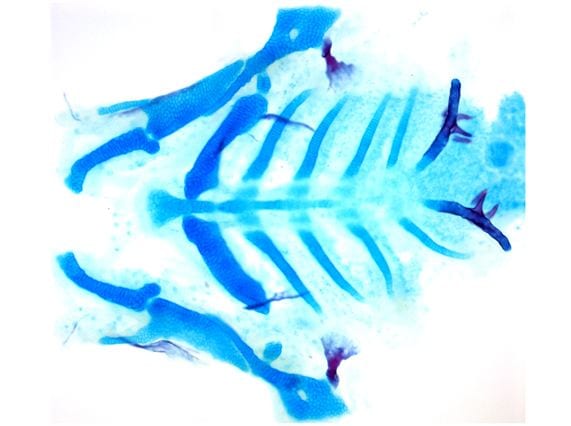
Dissection of the larval zebrafish skeleton shows facial cartilage (blue) and bone (red) from a ventral view. (Image courtesy of the Crump Lab) -

Lineage tracing shows endodermal contributions to the front of the head, including the pituitary. (Image courtesy of Peter Fabian) -

The larval zebrafish skeleton is shown with cartilage (blue) connected to muscles (white) through tendons (magenta).( Image courtesy of Olivia Chen) -
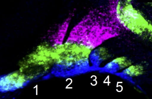
Pharyngeal arches house the neural crest-derived skeletal progenitors for the facial skeleton. During development, in situ RNA hybridization shows how they are separated into dorsal (magenta), intermediate (green), and ventral (blue) domains. (Image courtesy of the Crump Lab)