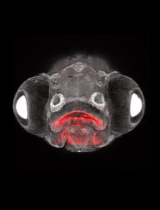
The pea-sized gland, nestled at the base of the brain, produces hormones that drive growth, aggression, sexual development and reproduction. For decades, the front lobe of the pituitary — where the hormones are made — was thought to be an evolutionary development that arose in vertebrates, along with the ear, the nose and the lens of the eye.
The widely accepted “new head hypothesis” holds that all of these body parts derive from a particular type of embryonic structure located in the ectoderm, or outermost layer of an embryo. Meanwhile, animals that have spinal cords but lack backbones, understood to represent an earlier evolutionary step, have a pituitary-like structure previously thought to have a distinct origin in the innermost embryonic layer, or endoderm.
In a paper published today in Science, USC researchers present evidence that, in some vertebrates, the endoderm also forms part of the pituitary’s front lobe — an idea that has been the subject of scientific controversy dating back more than 100 years. Findings from the study, which was supported by a major grant from the National Institutes of Health, suggest that the gland may have a longer evolutionary history than previously thought.
To read more, visit https://keck.usc.edu/pituitary-puzzle-gets-a-new-piece-revising-evolutionary-history/.
